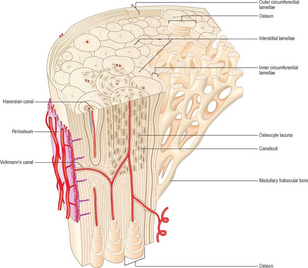
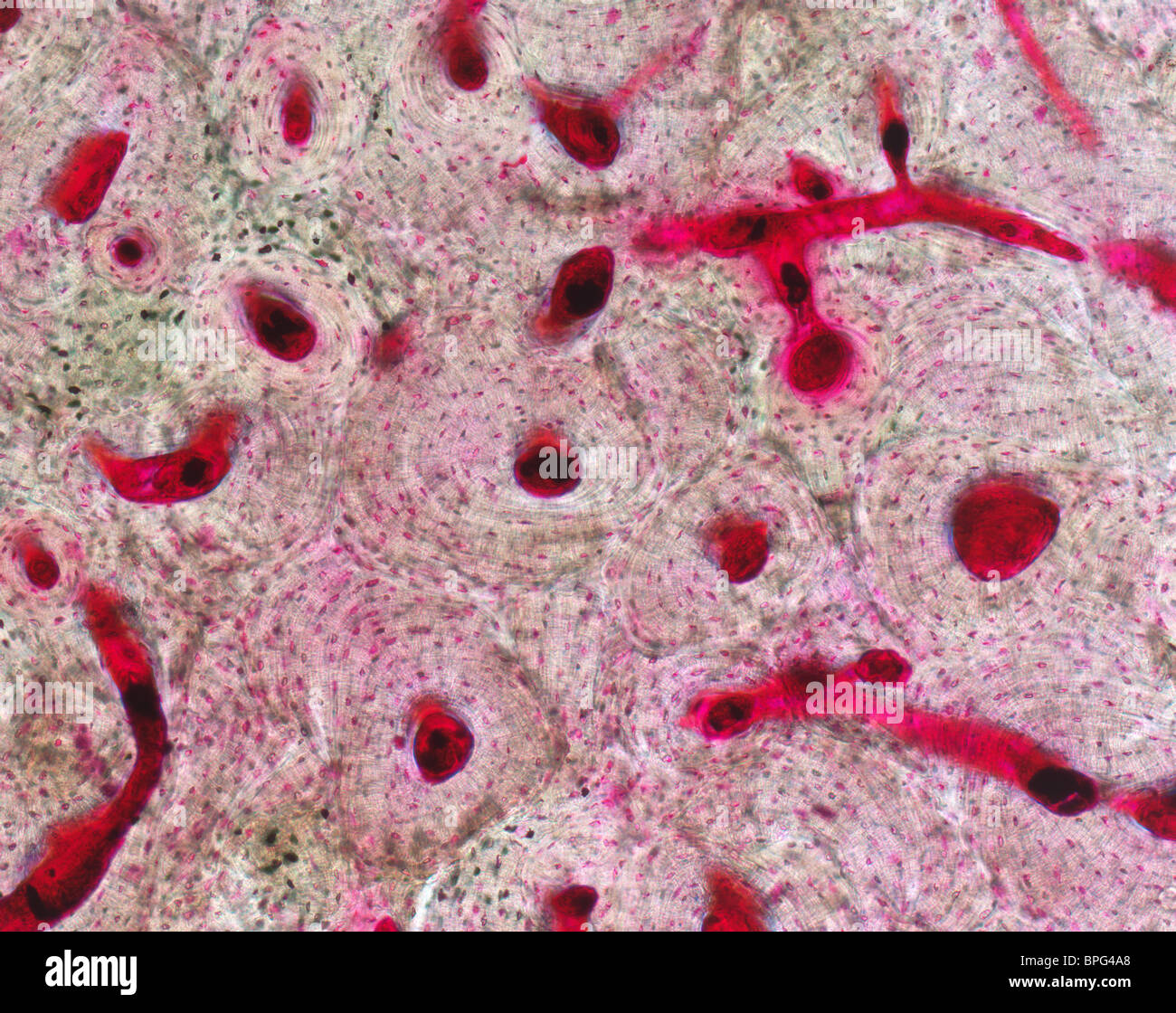
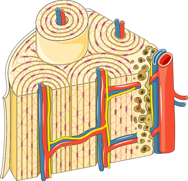
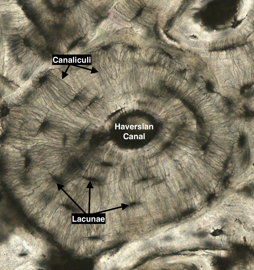
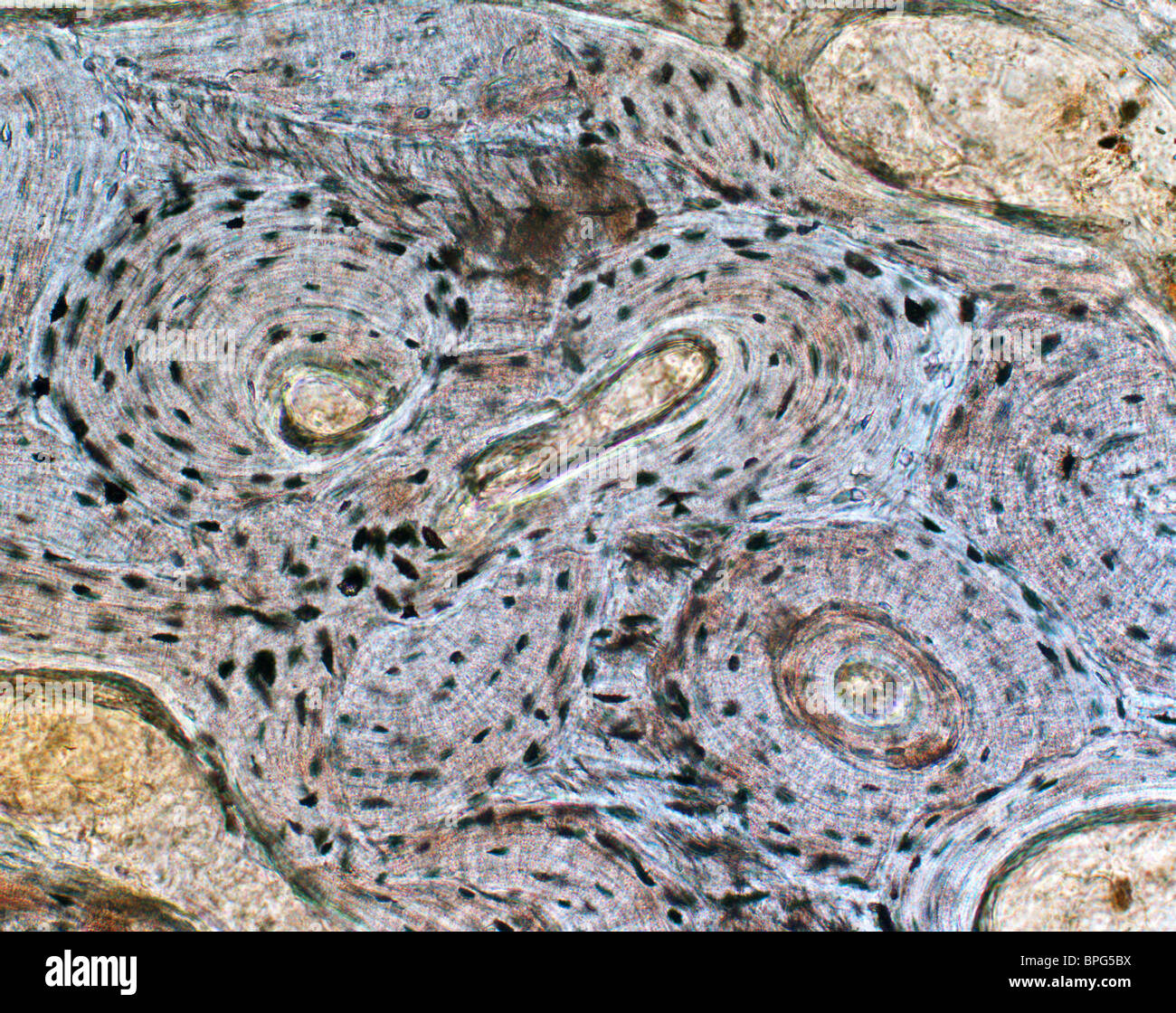
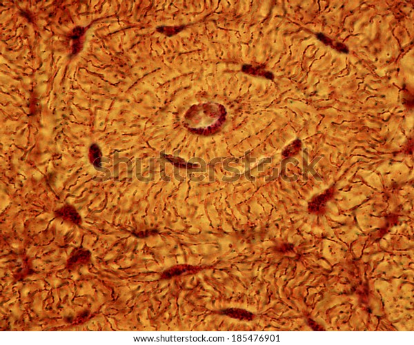
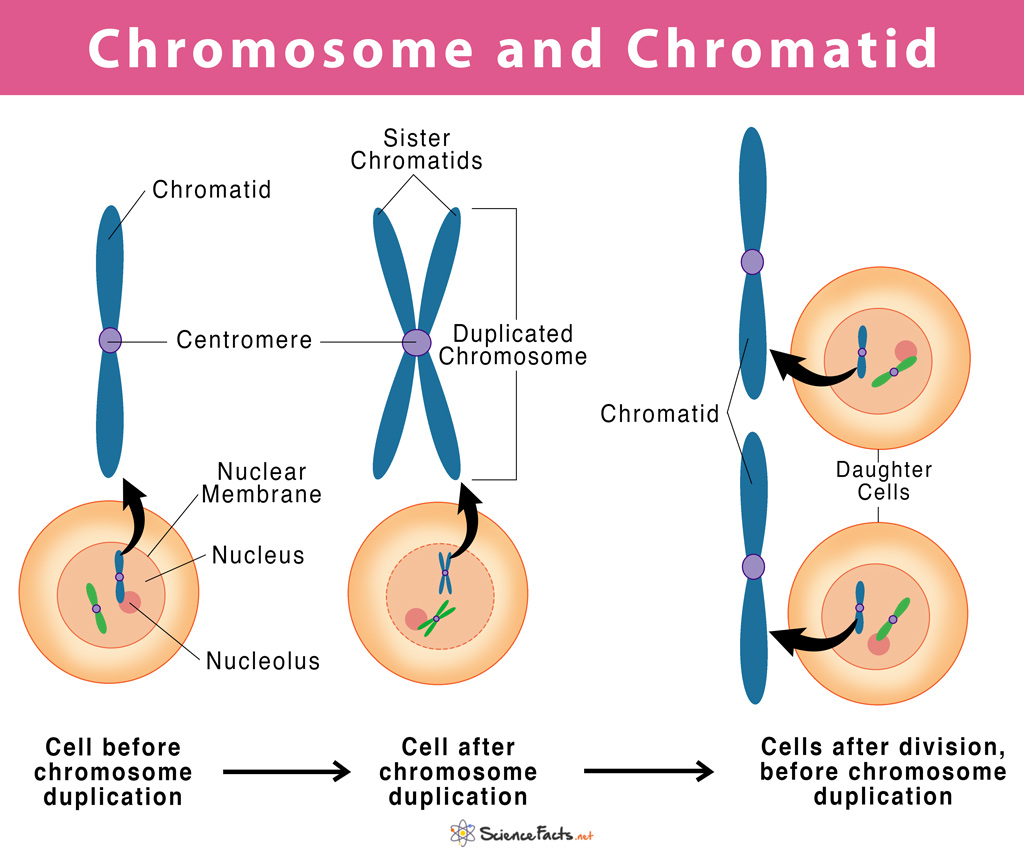
Albums 105+ Images concentric lamellae within an osteon are connected by lacunae. Superb

We present you with the fascinating concentric lamellae within an osteon are connected by lacunae. images from nghenhansu.edu.vn, thoughtfully compiled and presented. Explore further related images in the details provided below.

concentric lamellae within an osteon are connected by lacunae.

My Image 1
structure of compact bone Diagram | Quizlet
My Image 2
Illustration of compact bone showing the relationship between …
My Image 3
2: Compact bone organisation Top = The organisation of osteons and …
My Image 4
is the inner and outer circumferential lamellae is concentric or no
My Image 5
2 Cross-section of bone showing a cortical bone and spongy/cancellous …
My Image 6
This photo shows a model of an osteon. It points out the blood vessels …
My Image 7
Anatomy and Physiology 1 Chapter 6 Bone Tissue Flashcards | Quizlet
My Image 8
Regions and Structures of an Osteon Diagram | Quizlet
My Image 9
(A) Histological cross-section of cortical bone, showing osteon with …
My Image 10
Skeletal system: compact bone
My Image 11
Lacuna Anatomy
My Image 12
Correctly Label the Following Anatomical Parts of Osseous Tissue …
My Image 13
33.2C: Connective Tissues: Bone, Adipose, and Blood – Biology LibreTexts
My Image 14
Microscopic structure of Compact and Spongy bone tissue Diagram | Quizlet
My Image 15
Microscopic structure of human lamellar bone. 35 | Download Scientific …
My Image 16
lamellae anatomy bone
My Image 17
Lamellae Definition Anatomy – Anatomical Charts & Posters
My Image 18
Lacuna Definition and Examples – Biology Online Dictionary
My Image 19
Spongy And Compact Bone Diagram / Diagram Human Bone Anatomy Useful …
My Image 20
This pictures shows different views of compact bone. Important labels …
My Image 21
Osteon Diagram
My Image 22
6: Bone | Pocket Dentistry
My Image 23
Pin on Anatomy and Physiology
My Image 24
Osteon structure scheme. a = Haversian canal diameter; b = osteon …
My Image 25
Osteon Bone Haversian Blood Vessels | Art In Anatomy
My Image 26
Osseous Stracture Human Body : Human Body | General Science Quiz …
My Image 27
A. Secondary Osteon and B. Microscopic Structure of Mature Long Bone …
My Image 28
Bones: Structure and Types | Concise Medical Knowledge
My Image 29
1: a) Osteon, formed by bone lamellae with the haversian channel in the …
My Image 30
Bones at Liberty High School – StudyBlue | Bones, Anatomy and …
My Image 31
30 In The Diagram, Where Is The Osteon_ – Wiring Diagram Database
My Image 32
Osteon haversian system, lamellae, blood vessel continues…
My Image 33
Osteoblasts, Osteoclasts, Calcium, and Bone Remodeling | Owlcation
My Image 34
6 A single osteon. The osteon is drawn as if pulled out like a …
My Image 35
Solved Is it…. #N# a) canaliculi. b) lacuna. c) osteon. | Chegg.com
My Image 36
Compact Bone Diagram Lacunae : Bone Histology General Overview Compact …
My Image 37
Compact Bone Diagram Lacunae – Cartilage Bone Ossification The …
My Image 38
Secondary Osteon
My Image 39
Osteon Anatomy
My Image 40
Structure of an Osteon Diagram | Quizlet
My Image 41
Osteon Bone Haversian Lacunae Canaliculi | Art In Anatomy
My Image 42
Pin page
My Image 43
(A) Histological cross-section of cortical bone, showing osteon with …
My Image 44
Study Notes
My Image 45
Anatomy lab exam Tissues Flashcards | Easy Notecards
My Image 46
Functional anatomy of the musculoskeletal system | Basicmedical Key
My Image 47
Structure Of Compact Bone And The Osteon – sharedoc
My Image 48
Bone | Basicmedical Key
My Image 49
Light Micrograph Of An Osteon In Compact Bone Showing Osteocyte Lacunae …

LE MIGLIORI DIFESE D’EUROPA SU SORARE
My Image 50
What Is the Function of Compact Bone? (with pictures)
My Image 51
Compact bone tissue Diagram | Quizlet
My Image 52
Micrografía de una luz de hueso mostrando las laminillas concéntricas …
My Image 53
osteon – Liberal Dictionary
My Image 54
Histology Glossary: Histology – Compact Bone | Draw It to Know It
My Image 55
Cartilage/Bone/Muscle Histology Notes – Medical Histology – Jacobs …
My Image 56
Structure of Compact bone Diagram | Quizlet
My Image 57
Micrografía de una luz de hueso mostrando las laminillas concéntricas …
My Image 58
Light Micrograph Osteon Compact Bone Showing Stock Photo (Edit Now …
My Image 59
Activity 4: Examining the Microscopic Structure of Compact Bone …
My Image 60
A light micrograph of bone showing the concentric lamellae which …
My Image 61
6.4: Compact bone contains parallel osteons, and spongy bone contains …
My Image 62
Are there holes in bones in order to let blood cells out? If not, how …
My Image 63
A photomicrograph of an osteocyte embedded in its lacuna. Evolved from …
My Image 64
Structure and Function of the Musculoskeletal System | Basicmedical Key
My Image 65
Structure and Function of the Musculoskeletal System | Basicmedical Key
My Image 66
Hormonal Control of Calcium & Phosphate Metabolism & the Physiology of …
My Image 67
Overview of the osteocyte network within an osteon. a) 3D rendering of …
My Image 68
Solved T u engell lamelle Concentric of Interst n lamellal | Chegg.com
My Image 69
MCQs on Cell, Tissue, Skeleton System and Joint Vipin Vageria
My Image 70
[Solved] Label the following on the Ground compact bone slide 1 …
My Image 71
Difference Between Osteoblasts and Osteocytes | Compare the Difference …
My Image 72
Skeletal Tissues | Basicmedical Key
My Image 73
bone tissue pptx – dr.suhaila – Muhadharaty
My Image 74
14.2 Animal Primary Tissues – Concepts of Biology – 1st Canadian Edition
My Image 75
10 Concentric and interstitial lamellar bone (transverse section-ovine …
My Image 76
Compact Bone Diagram Pearson : Medullary Cavity Wikipedia / What is the …
My Image 77
Anatomy 312 > Brad Seebach > Flashcards > Exercise 6 – Histology: Bone …
My Image 78
Microscopic Anatomy (Bone Cells) – Structures of Bones
My Image 79
Print Chapter 6: Osseous Tissue and Bone Structure flashcards | Easy …
My Image 80
The Skeletal System ‹ OpenCurriculum
My Image 81
Solved Label the photomicrograph of compact bone. Osteocyte | Chegg.com
My Image 82
6: Describe the structure of the osteon in cortical bone AND 7: Explain …
My Image 83
A-C Ultrastructure of bone. A Light microscope image of a ground …
My Image 84
Human Skeleton – Skeletal System Function, Human Bones
My Image 85
Tecido conjuntivo: funções, tipos e matriz extracelular | Kenhub
My Image 86
6.3 Bone Structure — Anatomy and Physiology – Anatomy and Physiology …
My Image 87
Histology – Spongy Bone | Nursing notes, Osteology, Bones
My Image 88
In The Diagram Where Is The Osteon – Wiring Diagram
My Image 89
The lamellar units consist of concentric layers of elastic lamellae …
My Image 90
Solved EXERCISE 6 LAB REPORT Lamellae Canaliculi Central | Chegg.com
My Image 91
BIO201-Bone
My Image 92
Compact Bone Diagram Class 9 : Diffusion/Osmosis at University of …
My Image 93
Osteon of bone hi-res stock photography and images – Alamy
My Image 94
11 best Chapter 7 – Bone Tissue images on Pinterest | Mcgraw hill …
My Image 95
Schematic of osteon. A . Cross section. Indicated are Haversian canal …
My Image 96
Solved Osteon Lacunae Central canal Lamellae Osteons SEM X | Chegg.com
My Image 97
(a) Basic bone biology. In (a), (A) macroscopically, bone appears as …
My Image 98
Model of fibrillar orientation relative to the osteon axis. The …
My Image 99
Study Notes
My Image 100
2 Lamellae in a circularly polarized microscopy image of human cortical …
My Image 101
A light micrograph of bone showing the concentric lamellae which …
My Image 102
Compact Bone Diagram Lacunae / Structure of compact bone. | Download …
My Image 103
(PDF) A comparison of the microarchitecture of lower limb long bones …
My Image 104
Study Notes
My Image 105
bone tissue pptx – dr.suhaila – Muhadharaty
We sincerely appreciate your interest in the article about concentric lamellae within an osteon are connected by lacunae. at nghenhansu.edu.vn. Feel free to leave your comments, and don’t forget to check out more related articles below. We hope you find them fascinating and informative.

Similar Posts